Comparative lead: why localized modeling matters
When choosing preclinical platforms, investigators frequently contrast systemic xenografts with orthotopic, in situ lung cancer approaches; the comparative logic is straightforward and practical. In situ models reproduce native tumor microenvironment features with in vivo cell–stroma interactions that systemic implants cannot replicate, and this realism often yields superior histopathology and biomarker concordance. Laboratories that also maintain panels of metabolic disease models—metabolic disease models—find that methodological consistency across indications improves translational signal, because endpoints such as fibrosis or metabolic phenotype are evaluated with the same rigor and platform. This comparative perspective underpins the rest of the discussion.
Benefit 1 — More faithful tumor microenvironment and orthotopic behavior
In situ lung cancer models place malignant cells within the native pulmonary niche, preserving local extracellular matrix, resident immune cells, and vascular architecture. Such fidelity improves relevance for studies of invasion, angiogenesis, and immune checkpoint interactions. For pharmacokinetics and pharmacodynamics studies the tissue context changes drug distribution; therefore, orthotopic placement often yields more predictive efficacy signals than subcutaneous tumors. The practical implication is fewer false positives when advancing candidates to clinical evaluation.
Benefit 2 — Superior end‑point richness for biomarker and imaging work
Because the tumor evolves in the correct anatomical setting, investigators obtain richer endpoints: multiplex immunohistochemistry, longitudinal imaging, and spatial transcriptomics map more meaningful patterns. This advantage matters when a program depends on imaging biomarkers or spatially resolved gene expression to define mechanism of action. Studies using orthotopic models allow concurrent collection of bronchoalveolar lavage, precise tissue punches for histopathology, and circulating biomarkers, supporting robust phenotyping without sacrificing internal validity.
Benefit 3 — Better alignment with immune and stromal responses — practical caveat included
Immune cell recruitment and stromal activation differ dramatically by site; in situ lung tumors recruit alveolar macrophages and unique fibroblast subsets that shape treatment response. This alignment enhances studies of immunotherapy combinations and microenvironment‑targeted agents. A caution: in situ models demand more sophisticated surgical or injection techniques and careful animal welfare monitoring — procedural variability can confound results if not standardized. — Small operational lapses in implantation technique produce large downstream variance, and teams must train rigorously to avoid artifactual outcomes.
Benefit 4 — Translational consistency across disease models
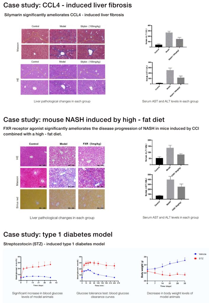
Programs that deploy both cancer and metabolic disease platforms gain a systems‑level view: inflammatory signaling, fibrosis progression, and metabolic dysregulation intersect in many human pathologies. For example, chemical‑induced liver injury models such as the ccl4 liver fibrosis model serve as a real‑world anchor for fibrotic pathway research and are commonly referenced in NIH-funded preclinical studies. Using orthotopic lung models alongside established fibrosis models improves comparative biology assessments and simplifies cross‑study biomarker translation.
Benefit 5 — Enhanced predictability for combination strategies and safety profiling
In situ tumors permit simultaneous assessment of local efficacy and organ‑specific toxicity, because the native milieu reveals off‑target interactions that ectopic sites may mask. Combination regimens that modulate vasculature, immune infiltration, or extracellular matrix can be tested with greater confidence. Teams concerned with safety find that orthotopic studies reduce late‑stage surprises, shortening iterative cycles between bench and preclinical validation.
Choosing the right model: three evaluation metrics
Metric 1: Biological fidelity — verify that the model reproduces key human features (stromal composition, relevant biomarkers, and expected histopathology). Metric 2: Operational reproducibility — mandate standardized implantation protocols, defined animal welfare monitoring intervals, and pre-specified phenotyping panels to reduce variance. Metric 3: Translational signal strength — require parallel readouts (imaging, circulating biomarkers, and tissue histology) so that efficacy and safety assessments triangulate toward a single conclusion. These rules guide selection of models and reduce downstream risk when moving toward IND‑enabling studies.
Conclusion
Comparative analysis shows that in situ lung cancer models provide measurable advantages in fidelity, endpoint richness, immunological relevance, translational alignment with models such as the ccl4 liver fibrosis model, and safety predictability — when teams commit to rigorous protocols. The three metrics above serve as golden rules for model selection. Jennio Biotech offers integrated platforms and standardized protocols that help implement these approaches smoothly — a practical solution for translational teams. —